Pelvic Anatomy : Normal Anatomy And Physiology Of The Female Pelvis Radiology Key / Surgical pelvic anatomy in gynecologic oncology.
byAdmin-
0
Pelvic Anatomy : Normal Anatomy And Physiology Of The Female Pelvis Radiology Key / Surgical pelvic anatomy in gynecologic oncology.. Branches of the internal iliac artery. Choose from 500 different sets of flashcards about pelvic anatomy on quizlet. Welcome to the valuemd albums. There are many organs that sit in the pelvis, including much of the urinary system, and lots of the male or female reproductive systems. Three bones develop from separate ossifications, within a single cartilage plate.
The pelvis (plural pelves or pelvises) is either the lower part of the trunk of the human body between the abdomen and the thighs (sometimes also called pelvic region of the trunk) or the skeleton embedded in it (sometimes also called bony pelvis, or pelvic skeleton). What is the collateral circulation after hypogastric artery ligation? Learn about pelvic anatomy with free interactive flashcards. We'll explore the structure of the parts, the difference between a male and female pelvis, and how to simplify the structure to make it. Pelvic anatomy includes both the male and female reproductive organs as well as important review pelvic anatomy with dr.
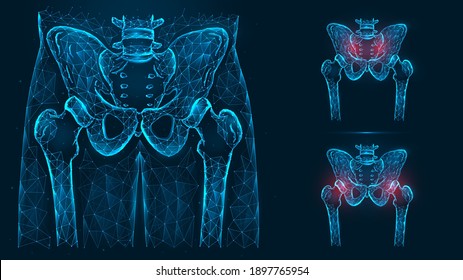
Pelvic Anatomy from www.bogg.com The pelvic brim (also known as the pelvic inlet) forms the superior margin of the lesser pelvis, separating it from the greater pelvis. Pelvic skeleton includes two hip bones, sacrum and coccyx. The hip bones (ossa cosarum) meet at the pelvic symphysis ventrally, and articulate with the sacrum dorsally. Supported along the lateral pelvic sidewalls by the ovarian ligaments (attaching to the posteriolateral. Anatomy of the human body for artists course. This mri pelvis cross sectional anatomy tool is absolutely free to use. Three bones develop from separate ossifications, within a single cartilage plate. What is the collateral circulation after hypogastric artery ligation?
It is composed of inlet, cavity, and outlet.
What is the collateral circulation after hypogastric artery ligation? Surgical pelvic anatomy in gynecologic oncology. Welcome to the valuemd albums. Prep for a quiz or learn for fun! Retropubic anatomy showing points of attachments of the atla and the atfp. Branches of the internal iliac artery. Choose from 500 different sets of flashcards about pelvic anatomy on quizlet. Interactive video showing normal female pelvic anatomy as seen by laparoscopy. The pelvic girdle consists of two symmetrical halves. Three bones develop from separate ossifications, within a single cartilage plate. Supported along the lateral pelvic sidewalls by the ovarian ligaments (attaching to the posteriolateral. The bony pelvis & gender differences in pelvic anatomy. Anatomy pelvis muscles pubococcygeus, puborectalis and iliococcygeus., pelvis nerve, the spinal nerves that arise from.
The pelvic brim (also known as the pelvic inlet) forms the superior margin of the lesser pelvis, separating it from the greater pelvis. It is composed of inlet, cavity, and outlet. Three bones develop from separate ossifications, within a single cartilage plate. Study pelvic anatomy using smart web & mobile flashcards created by top students, teachers, and professors. There are many organs that sit in the pelvis, including much of the urinary system, and lots of the male or female reproductive systems.
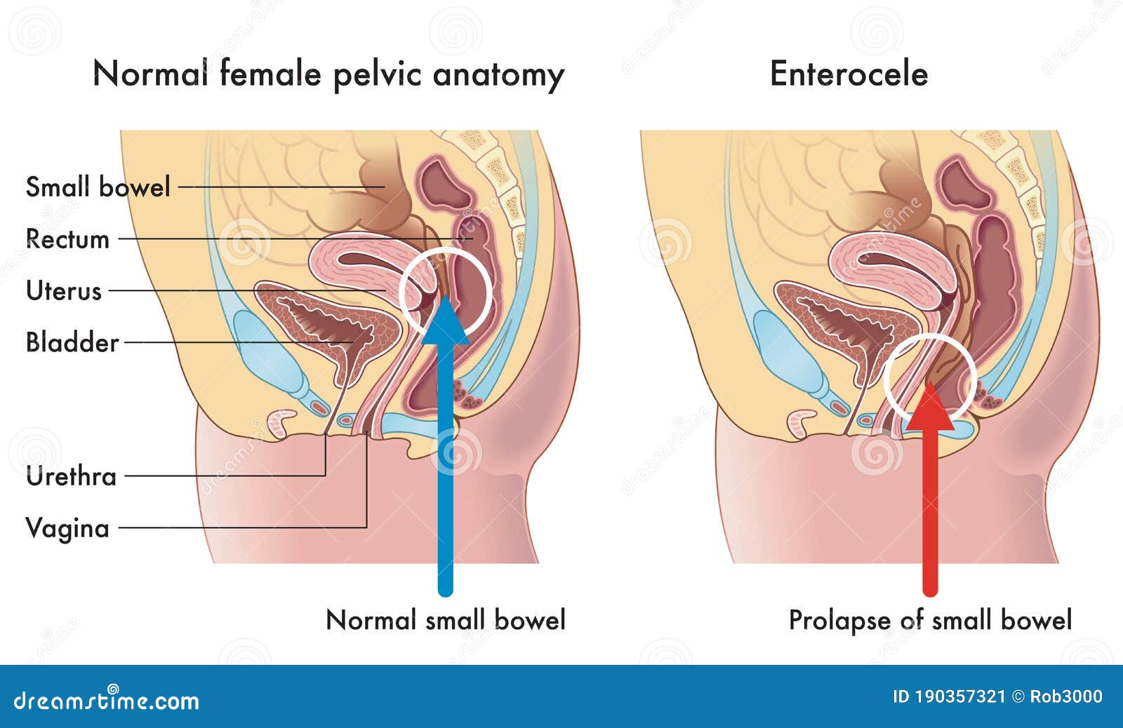
Pelvis Anatomy High Res Stock Images Shutterstock from image.shutterstock.com District i acog medical student education module 2011. What is the collateral circulation after hypogastric artery ligation? Anatomy of the human body for artists course. Learn about pelvic anatomy with free interactive flashcards. This article reviews normal pelvic anatomic findings during ultrasound and discusses how to obtain and measure these images. Female pelvis ppt by mayil rasamani 160332 views. Anatomy, pathologies and treatment options . Anatomy of pelvic organs uterus:
This section of the website will explain large and minute details of axial male pelvis cross sectional anatomy.
Anatomy of pelvic organs uterus: Abdominal and pelvic anatomy encompasses the anatomy of all structures of the abdominal and this anatomy section promotes the use of the terminologia anatomica, the international standard of. Register now and grab your free ultimate anatomy study guide! The pelvic brim (also known as the pelvic inlet) forms the superior margin of the lesser pelvis, separating it from the greater pelvis. Learn about pelvic anatomy with free interactive flashcards.
Female Pelvic Anatomy Stock Illustrations 453 Female Pelvic Anatomy Stock Illustrations Vectors Clipart Dreamstime from thumbs.dreamstime.com Welcome to the valuemd albums. Laparoscopic anatomy of the female pelvic region. Anatomy of the human body for artists course. Anatomy of pelvis & perineum by profgoodnewszion 75182 views. Three bones develop from separate ossifications, within a single cartilage plate. Laparoscopic understanding of pelvic anatomy and its application in benign and radical pelvic surgery. James pickering, associate professor of anatomy at the university of leeds. Above the pelvic brim and has no obstetric importance.
District i acog medical student education module 2011.
Above the pelvic brim and has no obstetric importance. Anatomy pelvis muscles pubococcygeus, puborectalis and iliococcygeus., pelvis nerve, the spinal nerves that arise from. Branches of the internal iliac artery. There are many organs that sit in the pelvis, including much of the urinary system, and lots of the male or female reproductive systems. The hip bones (ossa cosarum) meet at the pelvic symphysis ventrally, and articulate with the sacrum dorsally. Female pelvis ppt by mayil rasamani 160332 views. District i acog medical student education module 2011. Learn about pelvic anatomy with free interactive flashcards. This mri pelvis cross sectional anatomy tool is absolutely free to use. Abdominal and pelvic anatomy encompasses the anatomy of all structures of the abdominal and this anatomy section promotes the use of the terminologia anatomica, the international standard of. Laparoscopic anatomy of the female pelvic region. The bony pelvis & gender differences in pelvic anatomy. Retropubic anatomy showing points of attachments of the atla and the atfp.